Comparative Cellular Biogerontology

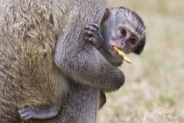
"Evolution is cleverer than you are." The best we can do, so far, within a species of mammals, is to postpone aging and extend lifespan by about 40% - 50%, using diets, drugs, or single gene mutations. But evolution, with more time and resources, has been able to construct species of mammals with maximum lifespans as long as ours (122 years) or whales (~200 years), using the same body plans and cellular toolkit that postpone aging for only 3 years in mice, 10 years in dogs, 20 years in horses, and about 30 years in chimpanzees. The most fundamental question in biogerontology is one of the simplest to frame: How does Nature produce long-lived species from short-lived progenitors? Evolution of longevity, i.e. evolution of mechanisms that postpone or buffer the effects of time on cell and tissue function, occurs repeatedly within the mammalian radiation, creating long-lived porcupines, humans, whales, bats, elephants, and mole-rats. Does Nature always use the same tricks, or a subset of them, to slow the aging process when doing so is adaptive? We hope that learning the answer to this question may help point the way to strategies that can further slow aging in people.
Our approach has been to collect skin-derived fibroblast cell line from many species of birds, rodents, primates, and “other” mammals (including bats, carnivores, shrews, etc.), and then to evaluate their traits in culture to see what properties are characteristic of cells from long-lived species. We hope in this way to identify cellular pathways that might also slow aging within a single species (mice, people) and give us clues about drugs that might use these pathways to slow aging and prevent late-life disease and disability. Our data across species, in culture, have so far shown consistent changes in (a) cellular resistance to many kinds of lethal injury, such as oxidation, UV light, DNA damage, and heavy metals; (b) the pace of activation of enzymes that mitigate cell stress; (c) ability to remove oxidized proteins in cells exposed to peroxide; (d) structure of the proteasome, a multi-protein enzyme complex that degrades damaged proteins; and (e) levels of an antioxidant enzyme, thioredoxin reductase 2, in mitochondria. See [link] for some of our recent papers in this area.
Our approach has been to collect skin-derived fibroblast cell line from many species of birds, rodents, primates, and “other” mammals (including bats, carnivores, shrews, etc.), and then to evaluate their traits in culture to see what properties are characteristic of cells from long-lived species. We hope in this way to identify cellular pathways that might also slow aging within a single species (mice, people) and give us clues about drugs that might use these pathways to slow aging and prevent late-life disease and disability. Our data across species, in culture, have so far shown consistent changes in (a) cellular resistance to many kinds of lethal injury, such as oxidation, UV light, DNA damage, and heavy metals; (b) the pace of activation of enzymes that mitigate cell stress; (c) ability to remove oxidized proteins in cells exposed to peroxide; (d) structure of the proteasome, a multi-protein enzyme complex that degrades damaged proteins; and (e) levels of an antioxidant enzyme, thioredoxin reductase 2, in mitochondria. See [link] for some of our recent papers in this area.